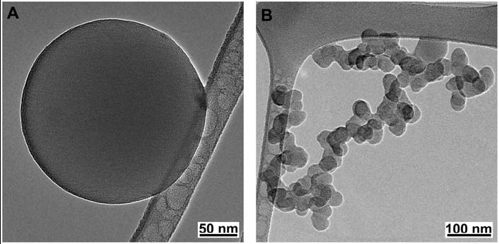
The first step in this project is to identify how many types of carbonaceous aerosols. From TEM, HRTEM and EDS, the morphology, structure and composition of the carbanecous aerosol particles can be determined. Up to now, we have found several typical carbonaceous aerosols from the samples collected from Yellow Sea region in East Asia. Their TEM images are shown following. (a) chain-like structured small spherule soot, (b) large spherule soot aggregate, (c) brown carbon sphere, and (d) cenosphere. We studied the concentration of these types of carbonaceous aerosol. The most important one is brown carbon and black carbon aerosol. Details are in our discussion paper published on Atmospheric Chemistry and Physics Discussion doi:10.5194/acpd-12-32945-2012
The EELS of brown carbon and black carbon shows different features. The π plasmon peak at around 5 eV of brown carbon is very broad, while it is much sharp for black carbon. We have published this result on Proceeding of Microscopy & Microanalysis 2012.
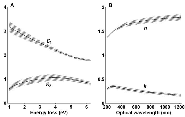
We studied the method to derive optical properties from their EELS data. A manuscript about the detailed processing is under preparation. Refractive index of brown carbon is shown below (Published on Science).
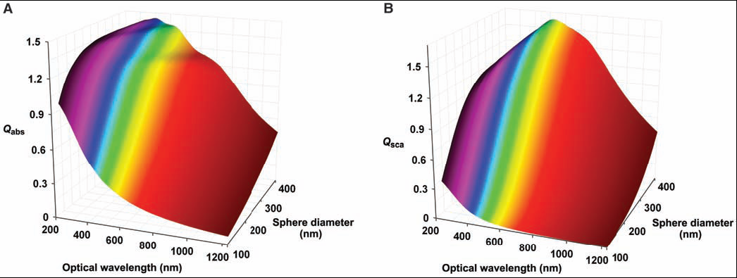
Once the optical properties of the carbonaceous aerosols are determined, their contributions in light absorption/scattering can be estimated. Here is showing the absorption efficiency of brown carbon spheres by Mie theory
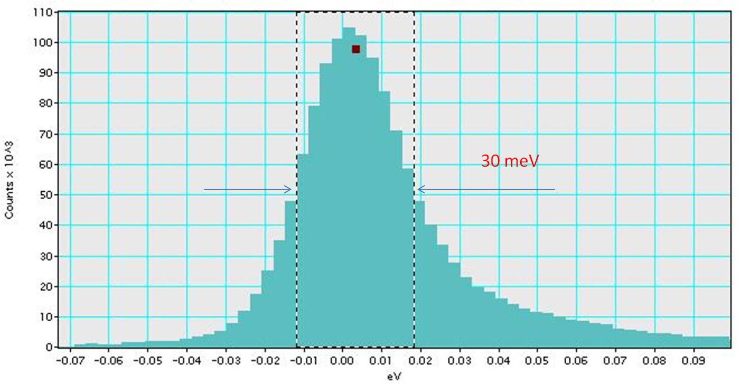
In principle, the dielectric function could be obtained from EELS in the full energy range. However, the limit of energy resolution is normally restrict the range of the derived dielectric function. The above work was done on monochromated TEAM 1 at National Center for Electron Microscopy. Now, ASU has a monochromated Nion UltraSTEM 100 which has shown a working energy resolution about 30 meV as shown following. We are working on this new microscope to get good results.